
1. What is Obstetric Ultrasound?
-
Obstetric ultrasound (also called prenatal ultrasound or obstetric sonography) uses high-frequency sound waves (via a transducer) to obtain images of the pregnant uterus, the embryo/fetus, placenta, amniotic fluid and maternal pelvic anatomy.
-
It does not use ionising radiation and is considered very safe when used appropriately.
-
It is widely used in obstetrics for dating the pregnancy, assessing viability, anatomy, growth, and monitoring for complications.
2. Techniques & Modalities

Approaches
-
Transabdominal ultrasound: The usual approach for many gestational ages — a curvilinear (or sector) probe is placed on the mother’s abdomen with ultrasound gel. Useful once uterus is large enough.
-
Transvaginal ultrasound: Especially useful in early pregnancy (e.g., 10 weeks) or when higher resolution of the cervix or lower uterine segment is needed.
Advanced/Additional Modes
-
2D (B-mode): Standard grayscale imaging.
-
Doppler ultrasound: To assess blood flow (e.g., umbilical artery, middle cerebral artery, fetal heart) — helpful in growth restriction or high-risk pregnancies.
-
3D and 4D ultrasound: (Three-dimensional imaging, real-time four-dimensional) used increasingly for fetal anatomy or research/parental imaging but not always part of routine scanning protocols.
3. Common Indications & What We Look For
Key Uses
-
Confirm the presence of a live intrauterine pregnancy (embryo/fetal heart motion) and exclude ectopic pregnancy.
-
Estimate gestational age (dating) and set an estimated due date (EDD).
-
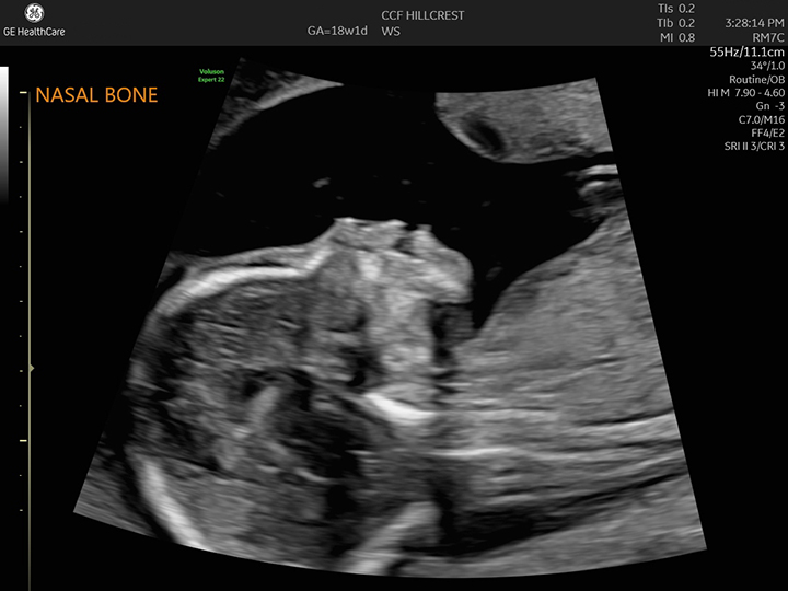
Assess fetal anatomy (especially in the second trimester, e.g., ~18-22 weeks) to identify structural anomalies.
-
Monitor fetal growth, amniotic fluid volume, placental location and maturity, fetal presentation (cephalic, breech), multiple pregnancies.
-
Evaluate maternal pelvic structures (uterus, cervix, ovaries) when relevant (e.g., shortened cervix, placenta previa).
Timing of Routine Scans
-
Many guidelines recommend at least two scans in an uncomplicated pregnancy: one in the first trimester (or early second) for dating and viability, and an anatomy scan in the mid-second trimester (≈18-20 weeks).
-
More frequent or specialized scans may be used in high-risk pregnancies.
4. Key Measurements and Fetal Biometry

Some of the important biometry measurements include:
-
Biparietal diameter (BPD) — measurement across fetal head.
-
Head circumference (HC)
-
Abdominal circumference (AC)
-
Femur length (FL)
These are used to estimate fetal weight, growth trajectory, gestational age.
5. Limitations & Pitfalls
-
Accuracy depends on operator skill, fetal position, maternal habitus (e.g., obesity), gestational age.
-
Some anomalies may not be detectable with ultrasound (depends on severity, timing, resolution).
-
Although considered safe, non-medical use (e.g., “keepsake” videos) is discouraged by authorities.
6. Safety Considerations
-
Diagnostic obstetric ultrasound has been extensively studied and no known harmful effects have been conclusively demonstrated when used properly.
-
The principle of ALARA (“as low as reasonably achievable”) applies — minimal exposure for diagnostic need.
-
Doppler mode involves higher acoustic output, so its use should be justified.
7. Practical Tips for the Sonographer/Clinician
-
Ensure the patient is comfortable; for transabdominal scans a partially full bladder may help in early pregnancy for better image window.
-
Use appropriate probe (curvilinear for abdomen; endovaginal for transvaginal).
-
Optimize image settings: depth, gain, focus, probe orientation.
-
Always document standard views and measurements; include fetal heart rate, placental location, amniotic fluid index (AFI) or deepest vertical pocket.
-
Correlate with clinical context (LMP, prior scans, risk factors).
-
Communicate findings clearly and document any abnormalities or concerns for further referral.
8. Summary
Ultrasound in obstetrics is an indispensable, safe, non-invasive imaging modality that allows assessment of fetal viability, growth, anatomy, and maternal structures. As a medical student (which I understand you are), mastering the common indications, standard planes and measurements will greatly aid your clinical competence in obstetrics and fetal imaging.

