0 Items
Wednesday, 22 October 2025 16:39

Kidney, Ureter and Bladder (KUB) Ultrasound

Written by
Rate this item
(2 votes)

Normal Ultrasound Appearance of KUB

Kidneys

https://www.researchgate.net/publication/344288950/figure/fig2/AS%3A943265876545540%401601903518372/Kidney-ultrasound-demonstrating-a-longitudinal-view-of-normalsized-kidneys-and-a-shape.png
https://i.ytimg.com/vi/JqJtnmPU8bM/maxresdefault.jpg
https://www.renalfellow.org/wp-content/uploads/2019/02/NormalPOCUS.png
POCUS Gallery - Renal Fellow Network
Ultrasound of the Upper Urinary Tract | Radiology Key
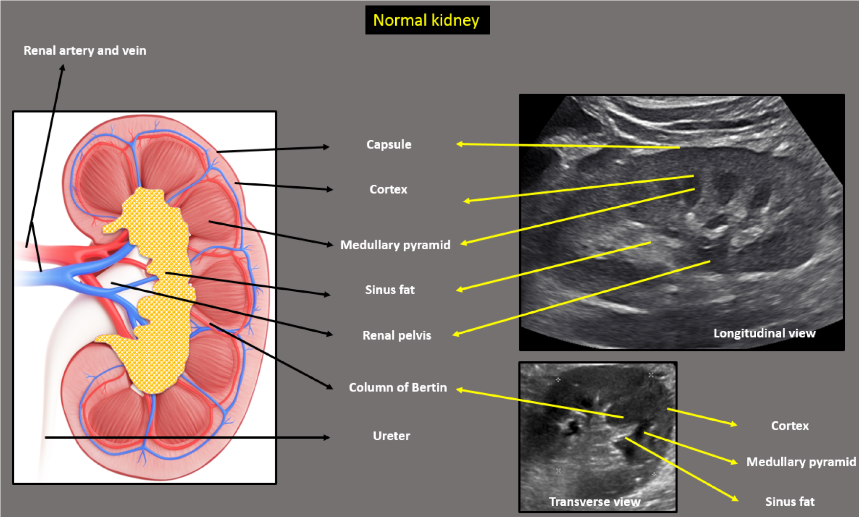
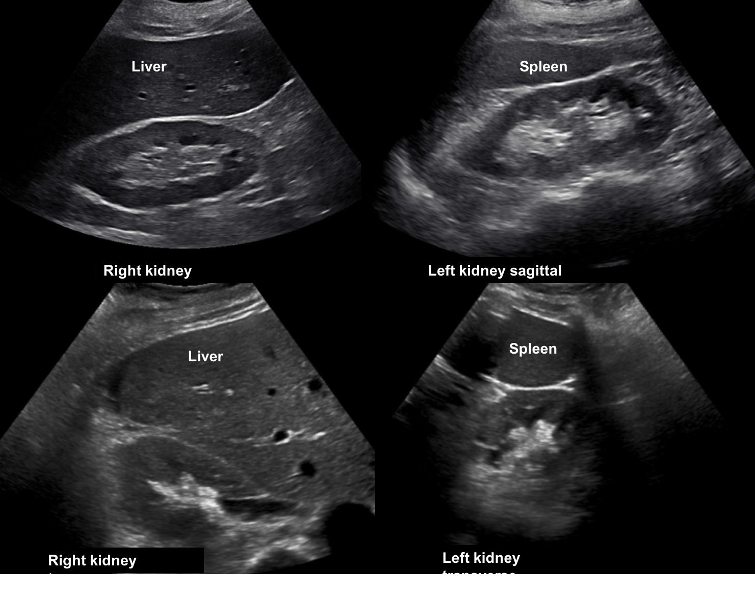
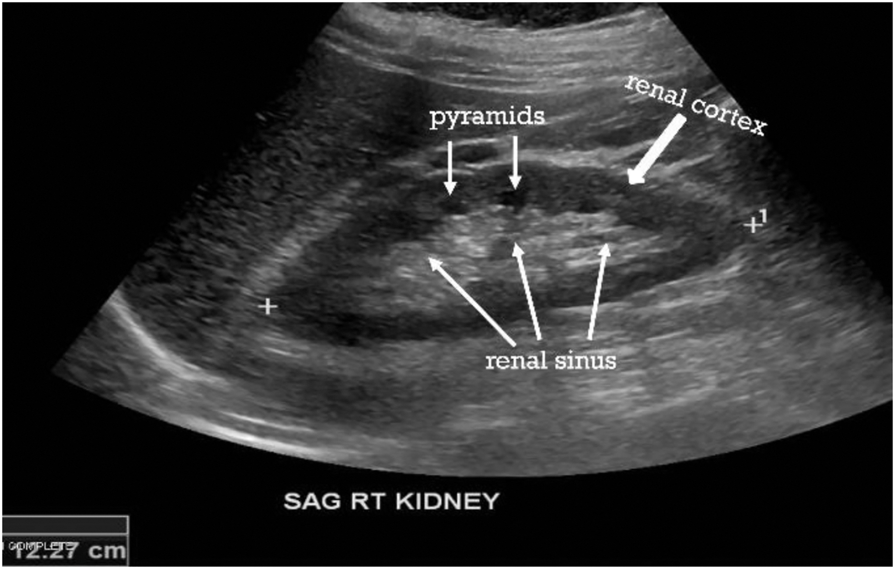
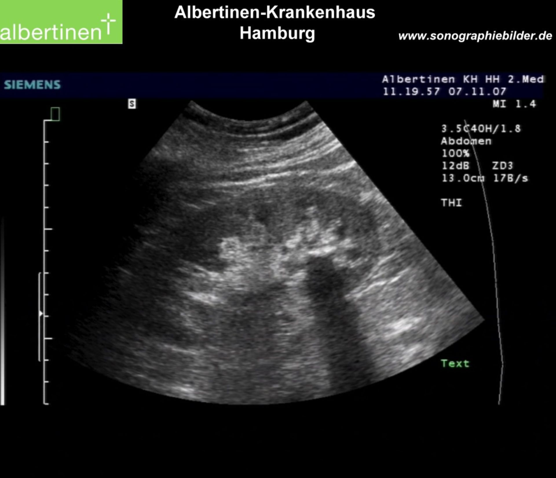
Kidneys are bean-shaped organs located in the retroperitoneum; on ultrasound you should appreciate the renal capsule (a thin hyperechoic line) and cortex and medulla differentiation. 
  • On longitudinal view, the kidney length in adults is typically ~9-12 cm (though varies by body size). 

  • The renal cortex is usually of similar echogenicity or slightly less than the liver in a non-fatty patient; the medullary pyramids appear as hypoechoic triangular/rounded areas. PMC

  • The renal collecting system (calyces/pelvis) should not be prominently dilated in the normal non-obstructed kidney.

Ureters

  • Normally the ureters are not well visualised on standard ultrasound unless dilated. 

  • At the bladder trigone you may see the ureterovesical junction region, but seeing a full course of ureter is often challenging due to overlying bowel, fat or other structures.

Urinary Bladder
https://www.imv-imaging.com/media/9120/image-1-bladder-wall.jpg
https://www.imv-imaging.com/media/9117/image-4-bladder-sediment.jpg
https://pocus101.b-cdn.net/wp-content/uploads/2020/09/Bladder-Ultrasound-Female-Transverse.png
 
Bladder ultrasound. A) Normal transverse ultrasound of a full ...
 
Urinary bladder wall thickness: what is the number to remember ...
 
Urinary bladder wall thickness: what is the number to remember ...
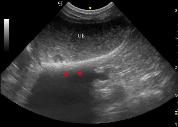
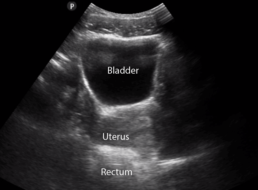
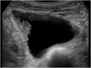
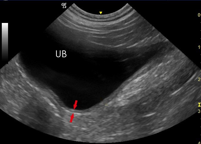
When adequately filled, the bladder appears as a well-defined anechoic (black) structure with thin, smooth walls. 
  • The bladder shape: in transverse view more rectangular/oval; in sagittal view a dome or triangle shape. 

  • The bladder wall should be thin (typically 4-5 mm) when fully distended and free of masses, debris or wall thickening.


🚨 Abnormal Ultrasound Appearances in KUB (Kidney/Ureter/Bladder)

Obstruction / Hydronephrosis
https://www.renalfellow.org/wp-content/uploads/2019/05/Figure-1-1024x585.jpg
https://images-provider.frontiersin.org/api/ipx/w%3D480%26f%3Dwebp/https%3A//www.frontiersin.org/files/Articles/538943/fped-08-00458-HTML/image_m/fped-08-00458-g004.jpg
https://www.meddean.luc.edu/lumen/meded/urology/ushydro3WLabels.jpg
6Hydronephrosis Grading Ultrasound | Kidney USG Scan Normal Vs Abnormal  Images | Grades 1, 2, 3 & 4
 
Kidney Ultrasound: HydronephrosisHydronephrosis appears on ultrasound as a dilated hypoechoic ...
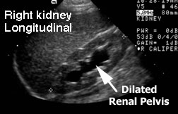
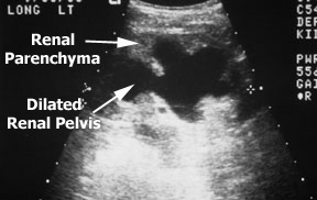
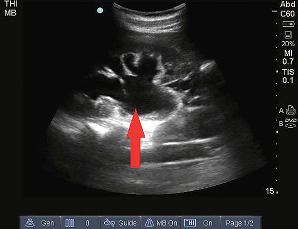
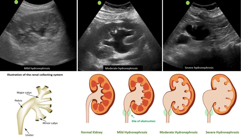
If urine outflow is blocked (e.g., stone, stricture), the collecting system dilates. On ultrasound you’ll see anechoic fluid in the renal pelvis and calyces, often described in grades (mild → severe). 
  • Secondary signs: loss of cortical-medullary differentiation, thinning of cortex, ureteral dilation (if visible) upstream of obstruction.

  • A key concept: Ureteral obstruction often first visible as hydronephrosis rather than seeing the ureter directly. 

Kidney Masses / Cysts

https://i0.wp.com/nephropocus.com/wp-content/uploads/2019/06/capture-2.jpg?fit=878%2C494&ssl=1
https://www.renalfellow.org/wp-content/uploads/2019/10/Figure-3.jpg
https://pub.mdpi-res.com/jcm/jcm-11-01112/article_deploy/html/images/jcm-11-01112-g001.png?1645695348=
6
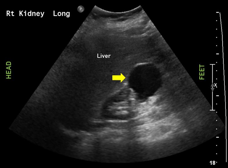
  • Simple cysts: anechoic, well-defined walls, posterior acoustic enhancement.

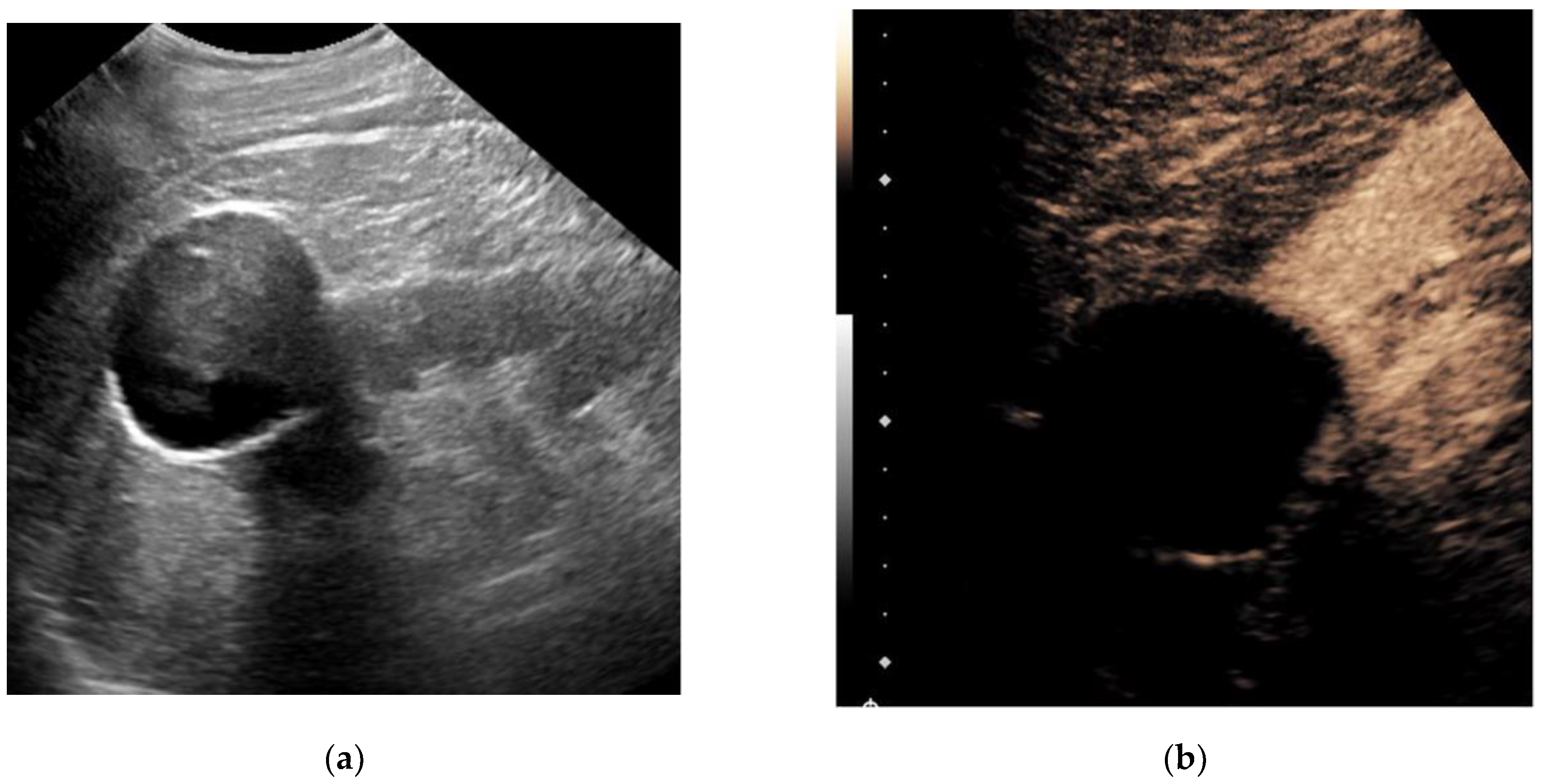
  • Complex masses: mixed echogenicity, irregular borders, may show internal vascularity. If found, further imaging (CT/MRI) is often required.

  • Polycystic kidney disease: multiple cysts replacing parenchyma, enlarged kidneys, loss of normal architecture.

Calculi / Stones

https://i0.wp.com/nephropocus.com/wp-content/uploads/2019/07/twitter-15.jpg?resize=878%2C494&ssl=1
https://dccdn.de/www.doccheck.com/data/kx/hk/6p/hc/vt/8b/nephrolithiasis-schallschatten-herold_lg.jpg
https://www.researchgate.net/publication/359172857/figure/fig2/AS%3A1132481680015360%401647016081095/USG-examination-of-the-bladder-showing-a-hyperechoic-shadow-indicating-a-foreign-body.ppm
6
  • Stones appear as hyperechoic (bright) foci often with posterior acoustic shadowing.

  • May see upstream dilatation (hydronephrosis). Stones in bladder may appear mobile/hyperechoic with shadow.

Bladder Abnormalities

https://prod-images-static.radiopaedia.org/images/48580315/536e85e24187d4f03627c69d66b679_gallery.jpeg
https://i0.wp.com/nephropocus.com/wp-content/uploads/2019/06/capture-4.jpg?fit=1200%2C675&ssl=1
https://www.ultrasoundcases.info/clients/ultrasoundcases/uploads/71414-Afbeelding1.jpg
6
  • Wall thickening may suggest cystitis, infiltration, chronic retention.

  • Bladder masses (tumours) may appear as echogenic intraluminal masses protruding into bladder, often with vascularity on Doppler.

  • Residual urine / urinary retention: large bladder filled with urine, may see debris, may see little change post-void.


📝 Summary Table: Normal vs Abnormal Features in KUB USG

Structure Normal Appearance Abnormal Appearance
Kidney Length ~9-12 cm, distinct cortex/medulla, non-dilated collecting system Enlarged/shrunken kidney, hydronephrosis, masses, cysts
Ureter Usually not visualised Dilated ureter, visible stone in ureter, ureteroceles
Bladder Anechoic lumen, thin smooth walls, well-filled Wall thickening, masses, stones, residual urine

✅ Why This Matters & Clinical Relevance

  • A well-performed KUB ultrasound is non-invasive, no radiation, and is a first-line imaging for many urinary tract conditions. 

  • It helps identify obstruction, which if untreated can lead to renal damage (e.g., hydronephrosis).

  • Detecting stones, cysts, and masses early can influence management significantly.

  • Bladder findings (stones, tumours, retention) may explain urinary symptoms (frequency, haematuria, retention).

Read 251 times Last modified on Wednesday, 22 October 2025 17:44
More in this category: « PERITONITIES CHEST ULTRASOUND »
Login to post comments

Overview

Health Africa is a tight-knit community of scientific, medical, and life sciences experts that produce and share the latest information, in a readable, understandable way. Also join our social nework group  Health Africa Conference for convenient communication. 

Get Support

For All Enquiries Contact Us

  • Mombasa, Kenya
  • +254 713 521 930
  • info@healthafricalibrary.co.ke

Login Form

Register to save more

Register an account today and get great deals, offers and customised products only for you.

  • Secure registration form
  • Your data on your hand, you can update anytime
  • Save your favourite products and access anytime
  • Order products easily.