
Normal Gallbladder Appearance

Key features:
-
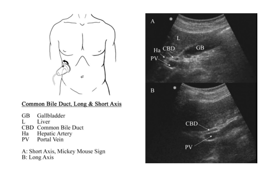
Pear-shaped, anechoic lumen (fluid filled) with clearly defined wall.
-
Wall thickness ≤ 3 mm when fully distended and in a fasted state.
-
Normal dimensions: about 7-10 cm in length, ~3-4 cm in transverse diameter in a fasted adult.
-
No intraluminal echogenic foci with shadowing, no wall irregularity or surrounding fluid.
Technique tips:
-
Have the patient fast (often 6-12 h) so the gallbladder is distended and easily imaged.
-
Use the liver as an acoustic window (right upper quadrant, subcostal or intercostal) to reduce bowel gas interference.
-
Scan both long axis and short axis, and fan through to cover fundus, body, and neck.
🚨 Abnormal Appearances & Common Pathologies
Below are key abnormal findings, what they look like on ultrasound, and what they may represent.
1. Gallstones (Cholelithiasis)

USG appearance:
-
Hyperechoic (bright) foci within the lumen with posterior acoustic shadowing (regardless of stone composition).
-
Mobility of stones when patient position changes (if not impacted).
-
A classic sign: the “WES sign” (Wall-Echo-Shadow) when gallbladder is packed with stones.
Clinical note: Stones are a common finding in gallbladder ultrasound and may warrant further management depending on symptoms.
2. Gallbladder Sludge

USG appearance:
-
Low-level, non-shadowing echoes layering in dependent portion of lumen.
-
May change with patient position but slower than free fluid.
-
No significant posterior shadowing (distinguishing from stones).
Clinical note: Sludge may represent biliary stasis; may predispose to stones or cholecystitis.
3. Acute Cholecystitis
USG appearance:
-
Wall thickening > 3 mm.
-
Distended gallbladder (often > 10 cm length) with gallstones impacted in neck or cystic duct.
-
Pericholecystic (surrounding) fluid or edema.
-
Positive sonographic Murphy’s sign: maximal pain when ultrasound probe presses over gallbladder.
Clinical note: Combined with clinical findings (RUQ pain, fever, WBC), these features support acute cholecystitis diagnosis.
4. Chronic Cholecystitis / Gallbladder Wall Thickening

USG appearance:
-
Diffuse or focal wall thickening (may mimic inflammation).
-
Features of Adenomyomatosis of gallbladder: Rokitansky-Aschoff sinuses (small intramural cystic spaces) and “comet tail” artefact.
Clinical note: Wall thickening may have multiple causes (inflammation, edema from systemic disease, neoplastic), so context is key.
5. Gallbladder Cancer / Mass-forming Lesions
USG appearance:
-
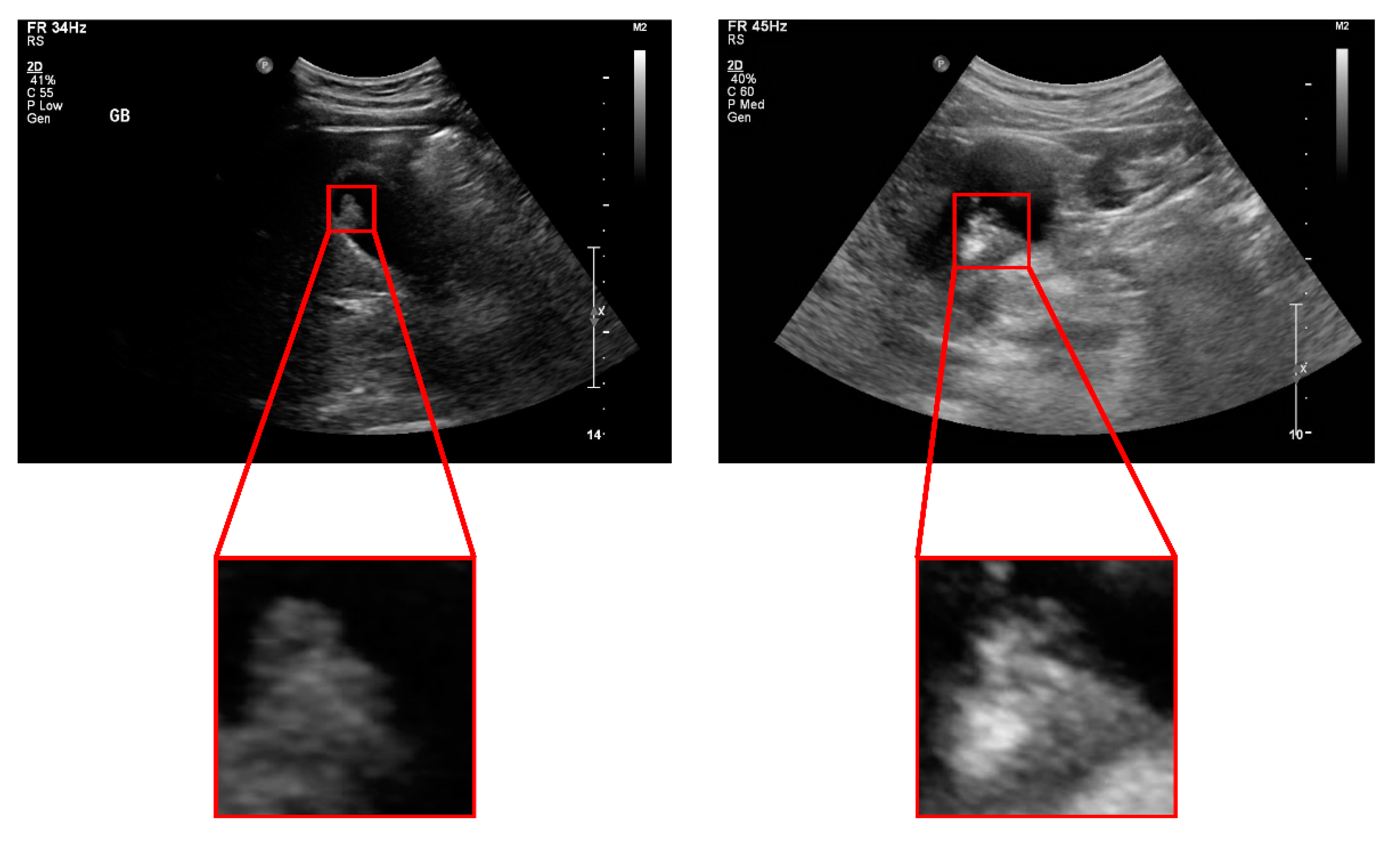
Focal intraluminal mass or thickened wall, often heterogeneous echotexture.
-
Irregular margins, possible calcifications, and may show invasion into liver parenchyma or adjacent structures.
-
Large gallbladder polyps (>10 mm) may raise suspicion for malignancy.
Clinical note: Suspicious features on ultrasound warrant further imaging (CT/MRI) and surgical consultation.
📝 Summary Table: Normal vs Abnormal Features
| Feature | Normal | Abnormal (Pathology) |
|---|---|---|
| Gallbladder shape & lumen | Pear-shaped, anechoic lumen | Distended with stones/sludge or collapsed/contracted |
| Wall thickness | ≤ 3 mm | > 3 mm (in inflammation) or significantly thick/mass-forming |
| Intraluminal echoes | None (just bile) | Hyperechoic stones with shadow, low-level sludge echoes |
| Mobility of intraluminal content | Free flow | Impacted stones, polyps fixed to wall |
| Surrounding fluid/edema | None | Pericholecystic fluid (in cholecystitis) |
| Mass or polyp presence | Absent | Intramural mass, large polyp, wall irregularity |

