
Chapter: Chest Ultrasound (POCUS)
1. Introduction
2. Anatomical & Sonographic Basics
2.1 Key Anatomy
-
Chest wall (skin → subcutaneous tissue → ribs → intercostal spaces)
-
The pleural interface: the parietal pleura (lining chest wall) and visceral pleura (covering lung)
-
The lung parenchyma filled with air (which usually impedes ultrasound beyond the pleura)
-
Diaphragm, liver (right side) and spleen (left side) serve as acoustic windows for inferior lung and pleural spaces.
2.2 Sonographic Landmarks
-
Pleural line: a bright, hyperechoic horizontal line just beneath the rib shadows, representing the interface of parietal & visceral pleura.
-
Rib shadows: When scanning longitudinally in an intercostal space, the ribs appear as bright lines with acoustic shadow behind. Useful to confirm correct probe positioning.
-
“Bat sign”: The two adjacent rib shadows appear like wings and the pleural line the body of the bat — useful to confirm correct intercostal window.
2.3 Ultrasound Artifacts & Their Meaning
-
A-lines: Horizontal, equidistant echogenic lines parallel to the pleural line. These arise due to reverberation in an aerated lung.
-
B-lines: Vertical, laser-like hyperechoic lines emanating from the pleural line, extending to the bottom of the screen, moving with lung sliding. They represent increased lung density/interstitial fluid.
-
Lung sliding: The shimmering motion of the pleural line as the visceral pleura glides over the parietal pleura during respiration. Its presence argues against pneumothorax in the scanned zone.
-
M-mode signs: In M-mode ultrasound, a normal sliding pleura produces the “seashore sign” (static tissue above pleura, granular below). In pneumothorax, absence of sliding gives a “barcode/stratosphere sign”.
3. Equipment & Scanning Technique
3.1 Probe Selection
-
Linear probe (5-10 MHz): Ideal for superficial structures (pleural line, pneumothorax detection) due to high resolution.
-
Curvilinear / phased array (2-5 MHz): Better penetration for deeper lung fields, pleural effusions, deeper consolidations.
3.2 Machine Settings & Positioning
-
Use a depth of ~4-8 cm for pleural line scanning; increase depth up to ~15 cm for deeper lung/effusion assessment.
-
Focus at pleural line. Reduce gain if artifacts (lines) are too bright or obscured.
-
Patient positions:
-
Anterior chest: supine or semi-recumbent.
-
Lateral/posterior: sitting or lateral decubitus, especially for posterior lung zones or pleural effusion.
-
3.3 Scanning Zones and Windows
-
Standard windows: anterior upper/lower, lateral upper/lower (each side) — sometimes posterior zones added.
-
The probe is placed perpendicular to the ribs in an intercostal space, with the marker toward the patient’s head (for longitudinal view) or toward the right side (for transverse).
-
Identify the rib shadows, pleural line, then assess artefacts (A-lines, B-lines), sliding, and deeper lung texture.
4. Normal Lung Ultrasound Appearance
Key features of a normal scan in an aerated lung:
-
Pleural line present, shimmering with respiration (lung sliding).
-
A-lines present (horizontal) beneath pleural line in many views, indicating air beneath.
-
At most 0-2 B-lines in any given intercostal zone (especially dependent zones) — more B-lines suggest pathology.
-
Curtain sign: At the lung base the moving lung “curtain” covers the diaphragm/liver or spleen with inspiration, arguing against large pleural effusion.
5. Pathological Findings & Their Ultrasound Patterns
5.1 Pneumothorax
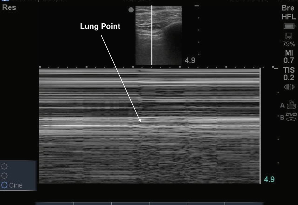
Ultrasound findings suggestive of pneumothorax:
-
Absence of lung sliding (pleural line static).
-
Absence of lung pulse (if ventilation suppressed but heart still generating pleural micro-movements).
-
Presence of A-lines only (air only) in the zone.
-
Lung point: Transition zone between sliding pleura and non-sliding pleura — highly specific for pneumothorax.
-
M-mode: “Barcode/stratosphere sign” instead of “seashore sign”.
5.2 Interstitial Syndrome / Pulmonary Edema
When there is increased lung density (fluid, fibrosis, interstitial thickening), typical signs include:
-
Multiple B-lines (≥ 3 in a zone) that move with respiration, extend to bottom of screen, erase A-lines.
-
The distribution and pattern matter: bilateral, diffuse B-lines → pulmonary edema; focal B-lines → pneumonia or localized interstitial involvement.
5.3 Consolidation & Pneumonia

Consolidated lung tissue (e.g., pneumonia) appears as tissue-like (“hepatized”) echotexture, often with:
-
Hypoechoic or heterogeneous echo pattern (“liver-like”).
-
Air bronchograms: small hyperechoic structures within consolidation that move (dynamic) with respiration → helps distinguish pneumonia from atelectasis.
-
The pleural line may appear thickened or interrupted, subpleural consolidations might be visible.
5.4 Pleural Effusion

Ultrasound is highly sensitive in detecting pleural fluid. Key features:
-
Anechoic or hypoechoic fluid collection above the diaphragm or between lung and chest wall.
-
Lung tissue may float or move within the fluid (if collapsed lung).
-
Complex effusion (exudate, empyema, hemothorax) may show septations, debris (“plankton sign”), or hematic layering (“hematocrit sign”).
5.5 Other Pathologies
-
Diaphragm dysfunction: assess diaphragmatic excursion or thickness.
-
Sub-pleural infarcts or pulmonary embolism: may show small, hypoechoic, wedge-shaped subpleural lesions.
-
ARDS: Mixed patterns with B-lines, spared zones, consolidations, pleural irregularities.
6. Focused Protocols & Clinical Application
6.1 The BLUE protocol for Acute Dyspnea
The BLUE protocol (Bedside Lung Ultrasound in Emergency) offers a structured scanning and interpretation algorithm for patients with acute respiratory failure.
Key profiles:
-
A-profile (A-lines + sliding) → suggests COPD/asthma in the right context.
-
A′-profile (A-lines without sliding) → suggests pneumothorax.
-
B-profile (≥3 B-lines bilaterally + sliding) → suggests pulmonary edema.
-
B′-profile (B-lines without sliding) → may indicate pneumonia.
-
C-profile (consolidation) → pneumonia, ARDS.
6.2 Trauma / eFAST Extension
In trauma settings, the chest POCUS is incorporated (into an eFAST scan) to detect pneumothorax and haemothorax rapidly.
6.3 Procedural Guidance
Ultrasound guidance for thoracentesis or chest tube insertion reduces complications by identifying the optimal fluid pocket and avoiding lung or diaphragm injury.
7. Advantages, Limitations & Pitfalls
7.1 Advantages
-
No ionizing radiation — safe for repeated use.
-
Bedside, real-time imaging allowing dynamic assessment.
-
High sensitivity for pleural effusion, consolidation near pleura, interstitial syndrome.
7.2 Limitations
-
Operator-dependent: image acquisition and interpretation need practice.
-
Air in the lung impedes ultrasound penetration → deeper lung parenchyma may not be visible.
-
Obesity, subcutaneous emphysema, dressings/tubes may limit access.
-
Certain patterns are non-specific (e.g., B-lines seen in edema, pneumonia, fibrosis) — always correlate clinically.
7.3 Pitfalls to Avoid
-
Mistaking Z-lines (short vertical artefacts not reaching bottom) for pathological B-lines.
-
Over-reliance on single view: pathology may be missed if scanning only anterior zones — posterior and lateral windows may reveal findings.
-
Assuming absence of sliding always = pneumothorax — pleurodesis, lung adhesions or large consolidation can also reduce sliding.
-
Misplacing probe (over rib rather than intercostal space) → poor view of pleural line.
8. Study Cases & Interpretation Examples
Case 1: Acute Dyspnea in Heart Failure
Scan shows bilateral diffuse B-lines, pleural sliding present → consistent with interstitial pulmonary edema. The presence of sliding helps exclude pneumothorax. (Supports clinical suspicion of heart failure exacerbation.)
Case 2: Post-Trauma Shortness of Breath
Anterior chest scan: absent lung sliding, only A-lines, and a lung-point seen laterally → suggests pneumothorax (small). Early detection allows prompt management.
Case 3: Fever, Cough and Hypoxia
Ultrasound shows subpleural consolidation with dynamic air bronchograms, plus some B-lines locally → consistent with pneumonia. Effusion unlikely (no large anechoic space).
Case 4: Fluid Accumulation in Pleural Space
Ultrasound identifies anechoic fluid above diaphragm with lung floating — guides thoracentesis site and shows layering (possible hemothorax if trauma).
9. Summary & Key Take-Home Points
-
Chest POCUS is a rapid, non-radiating, bedside imaging tool — highly useful for cardiopulmonary assessment.
-
Key sonographic landmarks: pleural line, rib shadows, A-lines, B-lines, lung sliding.
-
Normal aerated lung: pleural sliding + A-lines (few B-lines).
-
Pathologic patterns:
-
Pneumothorax → absent sliding, A-lines only, lung point.
-
Interstitial syndrome/edema → multiple B-lines.
-
Consolidation/pneumonia → tissue-like echotexture + air bronchograms.
-
Pleural effusion → anechoic/hypoechoic fluid above diaphragm.
-
-
Use structured protocols (e.g., BLUE) and include multiple zones (anterior, lateral, posterior) for thorough evaluation.
-
Always correlate with clinical presentation; ultrasound is a complement, not a replacement, for other assessments.
-
The biggest limitation is operator dependence — practice, image interpretation, and pattern recognition are essential.
10. Suggested Further Reading
-
Zadeh ES et al. “Lung Ultrasound and Pleural Artifacts: A Pictorial Review.”
-
“Lung Ultrasound: A Comprehensive Guide.” Stanford Medicine.
-
“POCUS Made Easy: Lung” — Life in the Fast Lane (LITFL).

