Introduction
Thyroid ultrasound (or thyroid sonography) is a non-invasive imaging technique that uses high-frequency sound waves to visualise the thyroid gland (lobes and isthmus), adjacent structures (such as trachea, carotid artery, jugular vein), and any abnormalities (nodules, cysts, thyroiditis, goitre).
It’s widely used in endocrinology and radiology for evaluation of thyroid size, texture, nodules, vascularity, and lymph-nodes in the neck.
Indications
-
Evaluation of a palpable thyroid nodule or neck mass.
-
Assessment of thyroid enlargement (goitre) or asymmetry of lobes.
-
Follow-up of known thyroid nodules (monitor growth or change in characteristics).
-
Guidance for fine-needle aspiration (FNA) of thyroid nodules or suspicious lymph nodes.
-
Evaluation of suspected thyroiditis (e.g., Hashimoto’s thyroiditis or Graves’ disease) or abnormal cervical lymph nodes.
Normal Sonographic Appearance

-
The thyroid gland typically has a homogeneous echotexture, meaning the internal echoes are uniform throughout the lobes and isthmus.
-
Echogenicity: medium to slightly high relative to surrounding neck muscles (strap muscles).
-
The lobes are connected by an isthmus; in transverse view it appears anterior to trachea.
-
Normal vascularity: visible but not overly dominant; Doppler flow should show scattered vessels, not an “inferno” pattern.
-
Measurements: Adult lobe typically length ~4–6 cm, AP 1.3–1.8 cm; isthmus thickness usually 1 cm.
Key Technical Protocol / Views

-
Patient lies supine, neck slightly extended. Gel applied over lower neck.
-
Transducer placed for transverse view at midline (isthmus) and then over lobes for transverse sweep (superior → middle → inferior).
-
Longitudinal (sagittal) view of each lobe (right and left) is obtained to measure craniocaudal length and AP dimension.
-
Colour or power Doppler is used to assess vascularity of gland/nodules.
-
Measurements: record length × width × height (or AP) of each lobe and isthmus. Document any nodules (size, echogenicity, margins, calcifications).
Sonographic Features of Abnormalities / Nodules
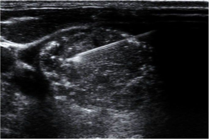
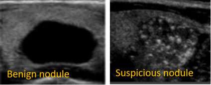
From the pictorial review by British Thyroid Association (U1–U5 classification) and other sources:
Benign features
-
Iso- or hyperechoic nodules with a halo (thin echogenic rim) often indicate benignity.
-
Coarse “comet-tail” artifacts and macro-calcifications may favour benign colloid nodules.
-
Spongiform appearance (microcystic change) is low suspicion.
Suspicious/malignant features
-
Hypoechoic nodules (especially markedly hypoechoic) compared to normal thyroid tissue.
-
Irregular margins, taller-than-wide shape on transverse image.
-
Micro-calcifications (tiny punctate echogenic foci) within a nodule: strongly suspicious for Papillary thyroid carcinoma.
-
Increased central vascularity on Doppler, or “thyroid inferno” pattern (seen in Graves’ disease).
-
Solid component, absence of halo, extrathyroidal extension, abnormal lymph nodes.
Thyroiditis / Diffuse disease
-
Heterogeneous gland echotexture, reduced echogenicity, increased vascularity.
-
In acute/subacute thyroiditis: may see hypoechoic areas, fluid collections.
How to Interpret & Report
When interpreting a thyroid ultrasound, consider:
-
Size, shape, and volume of each thyroid lobe & isthmus.
-
Echotexture and echogenicity of gland.
-
Presence of nodules: number, size, location (which lobe, upper/mid/lower), composition (solid, cystic, mixed), echogenicity, margin, calcifications, vascularity.
-
Adjacent structures: cysts, lymph nodes (cervical chain), extrathyroidal extension.
-
Colour-Doppler findings (vascularity).
-
Recommendation: Is follow-up required? FNA indicated? TI-RADS classification (or equivalent) may be used.
Advantages & Limitations
Advantages
-
Painless, no ionising radiation, real-time imaging.
-
High sensitivity for detection of nodules even when non-palpable.
-
Guides fine-needle aspiration safely.
Limitations
-
Operator-dependent; image quality varies with skill and equipment.
-
Ultrasound alone cannot definitively diagnose malignancy — features suggest risk, but cytology/histology often needed.
-
Some nodules deep, retrosternal or with calcified capsule may be difficult to visualise fully.
-
Acoustic shadowing (from calcifications), artefacts may obscure some regions.
Practical Tips for Performing the Exam
-
Use a high-frequency linear transducer (7–15 MHz) for superficial structure resolution.
-
Ask patient to tilt head slightly backward (neck extended) to improve access.
-
Apply sufficient gel, avoid air between probe and skin.
-
Sweep systematically: midline isthmus, right lobe (superior to inferior), left lobe similarly; obtain transverse and longitudinal views.
-
Measure nodules in three dimensions (length, width, depth) and record location (e.g., right lobe upper pole).
-
Use colour/power Doppler to assess vascularity especially if suspicion of thyroiditis or malignancy.
-
Compare with previous studies (if any) for size change.
-
Document any lymphadenopathy in the neck (especially lateral cervical).
Summary
Thyroid sonography is an essential imaging tool in evaluating thyroid pathology. Understanding the normal appearance, being familiar with the protocol views, recognising benign vs suspicious features, and appreciating its strengths and limitations will enable accurate reporting and better clinical decision-making.
A. Thyroid Cyst
-
Anechoic, well-circumscribed, posterior acoustic enhancement
📸
B. Colloid Nodule
-
Isoechoic/hyperechoic
-
Comet-tail artifacts (bright echoes)
📸
C. Thyroid Adenoma
-
Well-defined capsule, peripheral halo
-
Homogeneous, hypoechoic
📸
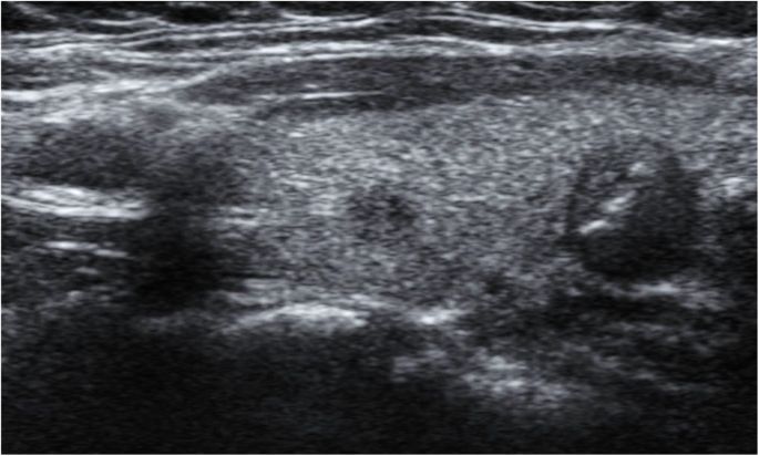
D. Papillary Carcinoma
-
Hypoechoic, irregular margins
-
Microcalcifications (tiny white dots)
-
Taller-than-wide shape
📸

E. Multinodular Goitre
-
Multiple nodules
-
Heterogeneous echotexture
📸

F. Hashimoto’s Thyroiditis
-
Diffusely hypoechoic, coarse echotexture
-
Increased vascularity
📸
G. Graves’ Disease
-
“Thyroid inferno” on color Doppler (diffuse flow)
📸

5️⃣ TI-RADS Classification (Simplified)
| TI-RADS Grade | Risk Level | Sonographic Findings | Management |
|---|---|---|---|
| 1 | Normal | Homogeneous gland | None |
| 2 | Benign | Spongiform or cystic | No FNA |
| 3 | Mild Suspicion | Isoechoic, smooth margin | Monitor |
| 4 | Moderate Suspicion | Hypoechoic, irregular margin | FNA if >1 cm |
| 5 | Highly Suspicious | Microcalcifications, taller-than-wide | FNA >1 cm, surgical review |
6️⃣ Reporting Template
Patient: Name / Age / Gender
Indication: Thyroid swelling, neck mass, follow-up, etc.
Technique: High-frequency linear probe (7–15 MHz), transverse and sagittal views.
Findings:
-
Right lobe: [Dimensions, echogenicity, nodules]
-
Left lobe: [Dimensions, echogenicity, nodules]
-
Isthmus: [Thickness, texture]
-
Vascularity: [Normal / Increased / Decreased]
-
Cervical lymph nodes: [Normal / Abnormal]
Impression:
-
Normal thyroid OR suggestive of [nodule type / thyroiditis / goitre].
-
TI-RADS score: [Insert grade].
-
Recommendation: FNA, repeat scan in 6–12 months, or referral to endocrinologist.
7️⃣ Case Studies (Educational)
Case 1: Benign Colloid Nodule
-
45-year-old female with anterior neck swelling.
-
Ultrasound: Isoechoic lesion, comet-tail artifact, peripheral halo.
-
Impression: TI-RADS 2 (Benign).
-
FNA: Colloid nodule.
Case 2: Papillary Thyroid Carcinoma
-
32-year-old female, right lobe hypoechoic nodule with microcalcifications, irregular margin.
-
TI-RADS 5
-
Histology: Papillary carcinoma confirmed.
Case 3: Hashimoto’s Thyroiditis
-
40-year-old female with fatigue and neck discomfort.
-
Ultrasound: Diffuse hypoechogenicity, coarse texture, increased vascularity.
-
Thyroid antibodies: Positive anti-TPO.
8️⃣ Practical Checklist (for Students and Sonographers)
| Step | Task |
|---|---|
| ✅ | Prepare high-frequency probe and patient in supine position |
| ✅ | Check gel, wipe air bubbles |
| ✅ | Start at isthmus (transverse), sweep both lobes |
| ✅ | Rotate probe for sagittal (longitudinal) view |
| ✅ | Measure each lobe (L×W×H) |
| ✅ | Record echogenicity, nodules, and vascularity |
| ✅ | Use Doppler for diffuse or focal flow |
| ✅ | Evaluate cervical lymph nodes |
| ✅ | Save images and measurements |
| ✅ | Complete structured report |
9️⃣ Summary Points
-
Thyroid ultrasound is the first-line imaging for all thyroid diseases.
-
Differentiates solid vs cystic and benign vs suspicious lesions.
-
Doppler helps assess vascularity in Graves’ or thyroiditis.
-
TI-RADS aids standardization and FNA decision-making.
-
Always correlate with clinical and biochemical findings (TSH, T3, T4, antibodies).
