PANCREAS
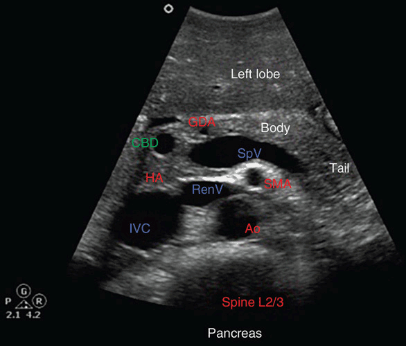
Normal anatomy & appearance
-
On ultrasound, the pancreas is located in the upper abdomen between the stomach (anteriorly) and the major vessels (posteriorly). A useful landmark is the Splenic Vein, which lies posterior to the body/tail of the pancreas.
-
The normal sonographic appearance: usually homogeneous echotexture, often isoechoic to slightly hyperechoic compared to the liver, though increasing fatty infiltration with age may make it more echogenic.
-
Normal duct diameter: The main pancreatic duct (MPD) is typically ≤ 2–3 mm. In the head region the common bile duct (CBD) is also visible (normal 4 mm) when well-seen.
Technique & scanning tips
-
Patient preparation: Fasting (6+ hours) helps reduce bowel gas and improves visualization.
-
Transducer selection: Use a curved array (e.g., 3–6 MHz) appropriate for penetration; in thinner patients higher frequency may be used.
-
Approach:
-
Start high in the epigastrum, transverse orientation, locate aorta/IVC as deep landmarks, then identify splenic vein and the gland above it.
-
To visualize the tail of the pancreas, a left inter-costal or subcostal approach using the spleen as acoustic window may be required.
-
Sweep through head → neck → body → tail, in both transverse and longitudinal planes.
-
Measurements (normal reference)
-
Sizes vary with age, habitus. Approximate normal values: head ≈ 35mm anterior–posterior, neck ~10-15mm, tail ~20mm (though wide variability).
-
Because pancreatic size correlates poorly with body size or age, the focus is often on morphology rather than exact measurements.
Common pathologies & ultrasound findings
-
Acute or chronic pancreatitis: gland enlargement, heterogeneity, increased echogenicity (in chronic), calcifications, duct dilatation.
-
Pancreatic cysts / pseudocysts: anechoic or complex fluid‐filled areas, often with history of pancreatitis.
-
Pancreatic adenocarcinoma or mass lesions: focal hypoechoic or heterogeneous masses, duct obstruction, vascular involvement.
-
Lipomatosis/fat infiltration: increased echogenicity of the gland.
Clinical tips
-
Always scan in more than one plane; a single view may miss pathology or mischaracterize.
-
Use surrounding vascular landmarks (splenic vein, SMV, aorta) to reliably locate the pancreas.
-
Be aware of limitations: bowel gas, patient habitus can degrade image quality.
-
Document both normal and abnormal sections: size, contour, echotexture, ductal changes, calcifications.
2. Spleen
Normal anatomy & appearance
-
The spleen lies in the left upper quadrant (LUQ), typically posterior, adjacent to diaphragm, stomach, and left kidney. It is intraperitoneal.
-
On ultrasound, normal spleen: homogeneous echotexture, often slightly more echogenic than the kidney.
-
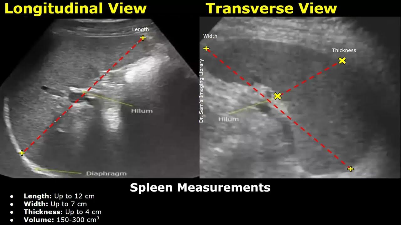
Normal size: According to measurement studies, upper limit of length ~12-15 cm in adults (varies with body size).
Technique & scanning tips
-
Patient preparation: Fasting may help reduce interfering bowel gas.
-
Approach: Subcostal or intercostal window in the left lateral decubitus or supine with left arm raised may improve access. The spleen can be visualized between ribs.
-
To measure: Place probe longitudinally (often between ribs), align with longest axis of spleen from upper pole (near diaphragm) to lower pole. Width (medial-lateral) and thickness (anterior–posterior) can be measured.
Measurements (normal reference)
-
Length: ~11–13 cm typical in many adults; however individual variation significant.
-
Thickness: 6 cm often used as cutoff when length borderline.
Common pathologies & ultrasound findings
-
Splenomegaly: enlarged spleen, rounded contour, may extend below left kidney.
-
Trauma / splenic laceration: irregular parenchymal defects, subcapsular hematoma, free fluid in splenorenal recess (“Koller’s pouch”).
-
Cysts / infarcts / abscesses: focal areas of altered echogenicity, fluid collections.
-
Accessory spleen (splenule): small extra nodules with similar echogenicity to spleen; common (≈10%)
Clinical tips
-
When measuring spleen, use consistent anatomical landmarks and scan plane to compare serially.
-
Assess for perisplenic fluid (especially in trauma).
-
Compare echogenicity of spleen to kidney (spleen often more echogenic than left kidney) as a quick check of texture.
-
Document any focal lesions, contour changes, surrounding fluid.
Summary Comparison Table
| Organ | Key Landmarks | Normal Appearance | Major Pathology Indicators |
|---|---|---|---|
| Pancreas | Splenic vein, aorta, IVC/SMV | Homogeneous, isoechoic/hyperechoic gland | Mass, cyst, duct dilatation, calcifications, heterogeneity |
| Spleen | Diaphragm, left kidney, ribs | Homogeneous, slightly more echogenic than kidney; length ~14 cm | Enlargement, focal lesions, perisplenic fluid |
